Horsetail Plant | Equisetum Arvense (Guide 2024)
Table of Contents
Equisetum arvense comprises about 25 species. It belongs to order Equisetales. They are worldwide in distribution; however, no species have been recorded from Australia and New Zealand. They grow in a variety of habitats.
The majority of the species are found in the north temperate zone. On the other hand, many species are tropical and found in South America and West Indies. The common Indian species are. E. debile, E. arvense and E. ramosissimum.
Certain species grow in ponds and marshy places, e.g., E. palustre; some species are shade-loving and grow in damp shady places, e.g., E. pratense whereas some other species grow in exposed conditions.
E. arvense is very widely distributed; E. debile is found along the banks of the rivers, canals, and pools in the Indian plains. Equisetum is commonly known as Horsetail Plant.
Classification Of Equisetum
The classification of plant Equisetum is given below along with the systematic position.
- Systematic Position: Pteridophyta
- Division: Arthrophyta.
- Class: Calamopsida
- Order: Equisetales
- Family: Equisetaceae
- Genus: Equisetum
Characteristic Features Of Order Equisetales
The sporophytes possess distinctly articulated stems. The ridges and furrows are present on the internodes of the stems.
The stems may or may not branch at the nodes. The branches are present in the whorls alternating with the leaves if branched. The leaves are scale-like, they form a toothed sheath around each node of the stem by their lateral fusion.
The stele is siphonostelic. It consists of a number of collateral vascular bundles in the internode which, however, join to each other forming a continuous ring at each node.
The sporangia are borne on the underside of peltate sporangiophores. The sporangiophores are arranged quite close to each other in terminal strobili or cone-like structures. The antherozoids are multiciliate.
Morphology Of Equisetum
Equisetum Habit
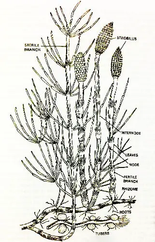
About all the species of Equisetum (Horsetail) are perennial. They bear much-branched, horizontal, creeping, and subterranean rhizomes which sometimes penetrate 3 or 4 feet deep in the soil.
The rhizomes bear erect and aerial branches. Some of the aerial branches are sterile and some are fertile. The plants vary in their height from species to species.
E. scirpoides is only a few inches high, whereas, on the other extreme a species of American tropics, i.e., E giganteum reaches up to 12 meters or 40 feet in height. However, its diameter is less than an inch.
The plant E. debile grows along the banks of the river and may attain a height of 10 to 15 feet when grown in damp and shady places. Its diameter is about one and a half centimeters.
Equisetum Stem
The rhizome of Equisetum (Horsetail) is horizontal, perennial, much-branched, subterranean, and penetrates in the soil up to 3 to 4 feet in many species.
It is spread over an area of 10 to 15 feet. It consists of well-differentiated nodes and internodes. At each node, there is a whorl of small, scale-like leaves united laterally to each other forming a brown sheath.
Alternating with each leaf of the sheath at each node of the rhizome, there is a branch primordium or bud.
These primordia may develop into aerial on subterranean branches immediately, or they remain dormant for a considerable long period and develop into new branches on the approach of favorable conditions.
Sometimes in certain species, e.g., Equisetum telmateia and Equisetum arvense the primordia develop into short rounded branches composed of only one internode. These rounded branches are known as tubers.
After being separated from the parent rhizome tubers develop into new plants. This is a method of vegetative propagation of the sporophyte generation.
The whorls of the much-branched adventitious roots have been given from the nodes of the rhizome.
Usually, the rhizomes bear two kinds of aerial branches-sterile and fertile, but they are still other species that bear either sterile or fertile branches.
The sterile branches are of green color and bear a whorl of lateral branches at each node. These branches are assimilatory in function and practically synthesize all the food required by the plant.
The lateral branches found on the primary branch also bear the whorl of branches which are comparatively smaller in size.
Each whorl bears an equal number of branches to that of leaves and alternates with the leaves. The sterile branches of a few species are unbranched, e.g., Equisetum hyemale.
The fertile branches are also found above the ground, they bear strobili at their apical ends. These branches arise first and shed their spores even before the appearance of the sterile branches.
In E. arvense the fertile shoots are unbranched and devoid of chlorophyll, and they die as soon as the spores are shed.
In certain other species, e.g., E. cylvaticum and E. pratense the strobili are thrown off after spore dissemination, and the fertile branch now acts as the sterile branch by developing lateral branches on it.
Simultaneously, chlorophyll is also developed in these branches. E. palustre bears three kinds of branches
- The sterile branches are deep green and much-branched.
- The fertile shoots are short-lived and devoid of chlorophyll.
- The intermediate type of branches are fertile, devoid of chlorophyll, and unbranched at first, but later on, the strobili are thrown off and the branch becomes green, branched, and persistent. This is a good example of division of labor in the (Horsetail) Equisetum plant.
As regards function, the rhizome acts as a storehouse of the food material and helps in anchoring plants to the soil. The aerial sterile branches are green, and therefore, they synthesize all the food required by the plant.
The fertile branches are without chlorophyll and bear the strobili at their apical ends. The internodes of both the rhizome and aerial branches are ribbed longitudinally. These ribs alternate with the leaves in the subtending node.
The ribs also alternate with the ribs of the successive node, in their position. The general structure of the stem is the same both in the rhizome and the aerial shoot, but it may be more clearly understood in the aerial shoots.
Equisetum Leaves
The leaves are arranged in whorls at each node of the aerial branches. They are simple, scaly, slender, uninured, brownish, and more or less fused at their bases laterally to form a sheath around the base of the internode.
Their apices are, however, free and pointed. The number of leaves in each whorl ranges from 3 to 40 from species to species.
At first, the leaves are green, but later on, they become brownish, dry, and scale-like. The mature leaves are protective in function. In certain other species, they become dead, dry, and scaly.
Equisetum Roots
In Horsetail all the roots are adventitious except the primary root of the young sporophyte. The roots arise in whorls from each node of the creeping, subterranean rhizome.
The roots may be many years together, but do not increase in their length. The roots do not arise directly but from the lateral branch primordia situated on the nodes of the rhizomes and main aerial shoot.
Anatomy Of Equisetum
Anatomy Of Equisetum Stem
To study the internal structure of the stem, the transverse and longitudinal sections passing through the nodes and internodes are required. This way the internodal and nodal anatomy of the aerial stem may be studied separately. It is as follows:
To study the anatomy of the node, one has to cut the aerial stem surface of the stem looks wavy, because of the presence of ridges and furrows.
Scouring Rush Definition
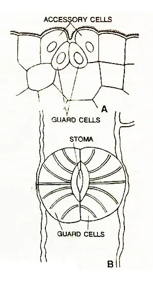
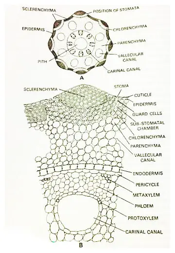
The stem is covered over by a single-layered epidermis interrupted by stomata situated in the grooves or furrows. The epidermis is always impregnated with a thick layer of silica.
The deposition of the silica on the epidermal layer gives a rough appearance to the stem, and therefore, the Equisetum plants are also known as ‘scouring rush or scouring rushes’.
The stomata are found in the grooves of the aerial shoots.
The development of the stomata is somewhat peculiar in the sense that the initial divide twice by successive longitudinal divisions and this way the two innermost cells develop into the guard cells.
Whereas the two outermost cells develop into accessory cells on being mature of the stomata.
The two accessory cells completely overarch the guard cells and the stoma.
In the majority of the species, e.g., E. hymeale, E. ramosissimum etc., the stomata are sunken, whereas in some other species, e.g., E. palustre, E. pratense, etc., they are situated on the surface of the epidermis.
The silica is deposited in the wall of guard cells in transverse radial bands.
Just underneath the epidermis, there is a broad cortex. The cortex consists of mechanical and assimilatory tissue. In the outer cortex, just beneath each of the ridges, there is a strand of sclerenchyma.
Usually, the sclerenchyma is restricted to the periphery but in Equisetum giganteum it extends inward to the endodermis. These columns of sclerenchyma are the chief mechanical elements of the shoot.
In addition to the above-mentioned sclerenchymatous columns, the sclerenchyma strands are also found in the furrows. Here, each strand is situated in between the curved strands of chlorenchyma.
The chlorenchyma possesses well-developed intercellular spaces. These spaces are very much distinct below the stomata. These chlorenchymatous bands are found beneath the sclerenchymatous strands situated underneath the ridges.
The ends of the chlorenchymatous bands touch the epidermis in the grooves. Since the leaves are small, scaly, and have a smaller number of chloroplasts the chloroenchyma of the cortex of the shoot is the major photosynthetic tissue.
The inner cortex is composed of thin-walled parenchyma with well-developed intercellular spaces. In this region of the cortex, the air spaces known as vallecular canals are also present.
Usually, vallecular canals are found opposite the furrows in the deeper tissue of the cortex. They are alternate to the vascular bundles. These canals are filled up with air.
The endodermis is the last layer of the cortex. In E. arvense, E. telmateia, E. palustre and certain other species, the endodermis surrounds the entire stele. In E. giganteum, E. limosum, and some other species, each vascular bundle is surrounded by a separate endodermal layer.
In other cases, e.g., E. hyemale, E. ramosissimum etc., there is a common internal endodermis inside the ring of bundles delimiting the pith, and a common outer endodermis found outside the ring of bundles.
Just beneath the endodermis, a single-layered pericycle is found. The pericycle is the outermost layer of the stele.
The vascular skeleton, i.e., the stele of Equisetum is siphonostelic. The separated vascular bundles are arranged in a ring.
The vascular bundles are situated opposite the ridges and alternate with the vallecular canals.
The number of bundles varies from species to species. In each internode, as many vascular bundles are found as there are leaves at the node.
The vascular bundles alternate with each other in their position in the successive nodes.
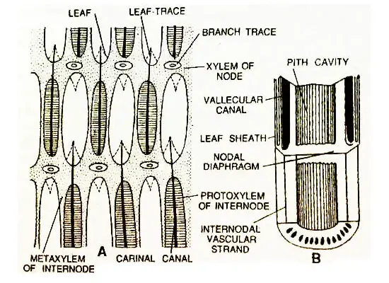
The internodal portions are longitudinally perforated, and each perforation runs the length of an internode.
The perforations in the internode are situated above the traces. These perforations are not branch gaps.
The vascular bundles are collateral type and resemble some extent to that monocotyledons. The vascular bundles contain both the metaxylem and protoxylem.
A carinal canal is developed in each bundle, because of the disintegration of the early-formed tracheids of the protoxylem during the elongation of the surrounding cells of the internode.
The remaining protoxylem elements are composed of a few tracheids. These protoxylic elements are found arranged to the margin of the carinal canal.
The metaxylem elements are found in two groups. The two groups are found arranged on the margin of the carinal canal towards the outside. The protoxylem lies in between the two groups of metaxylem.
The metaxylem elements are composed of reticulate, scalariform, or pitted tracheids. Sometimes spiral and annular tracheids are also found.
The protoxylem is endarch and as supported by Eames (1909) and Miss Barrat (1920) the development of the xylem is centrifugal.
The phloem is composed of sieve tubes and phloem parenchyma. The sieve plates may also be seen. The companion cells are not found.
The secondary growth is altogether absent. The central region of the stem is occupied by a hollow pith. The carinal canals are filled up with water.
Nodal Anatomy Of Equisetum
The alternate vascular bundles of the successive internodes are connected to each other by short branches and this way a continuous ring of the vascular cylinder is found in the node.
Eames (1909) reported that the bundles at the nodes do not have carinal canals. Here, the protoxylem elements are intact and completely occupy the lacuna or carinal canal.
At the node, the pith is not hollow and it forms a diaphragm separating the two successive internodes.
Rhizome
The anatomy of the rhizome is quite identical to that of the aerial shoot. However, the assimilatory tissue and the stomata are not found in the rhizome.
The mechanical tissue. i.e., sclerenchyma is poorly developed as compared to that of the aerial shoot. In E. arvense, the pith is solid, whereas, in certain other species, the pith and the vallecular canals are very much reduced.
Leaf (Anatomy)
The anatomy of the leaf is quite simple. The leaves are unnerved, this means that each leaf contains a single vascular bundle.
The vascular bundles of the leaf sheath are simple and collateral. The carinal canals are not found.
Individual bundles are surrounded by separate endodermal layers (endodermis).
The outer tissues of the leaf sheath are sclerenchymatous bands.
These bands of sclerenchyma pass up the leaf ridges and alternate the strips of chlorophyllous tissue associated with stomata.
Root (Anatomy)
The adventitious roots are borne at the nodes of the rhizome and aerial shoot. The anatomy of adventitious roots is quite simple.
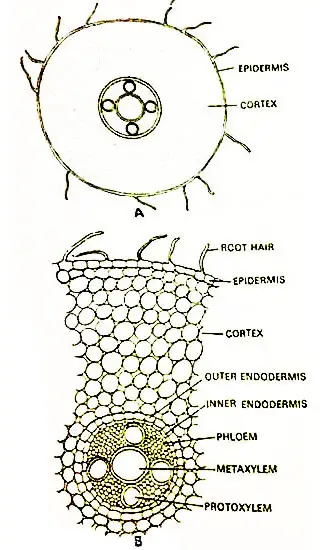
To study anatomy, a cross-section of the root is required. The outermost layer is known as the piliferous layer which bears unicellular hairs upon it.
Just beneath the piliferous layer, there is a multilayered cortex. In small roots, the cortex is divided into two zones.
The outer zone is composed of three to four-layered lignified exodermis.
The inner zone of the cortex consists of thin-walled parenchyma with well intercellular spaces.
The endodermis is two-celled in thickness. The inner layer of the endodermis acts as a pericycle. However, the pericycle is absent.
The lateral roots originate from the inner layer of the endodermis. In between the two layers of the endodermis, intercellular spaces are found. The Casparian is strictly found in the outer layer of the endodermis.
The stele of the root varies in nature from species to species. It is triarchy to hex-arch. There is a big central metaxylem element surrounded by three to six narrow points of protoxylem. Each protoxylem point is represented by a single tracheid.
The tracheids of xylem elements are spirally thickened. The angles between the protoxylem points are completely filled with phloem. The phloem is composed of phloem parenchyma and sieve tubes.
The Apical Growth
The apical growth of the main aerial shoot and its laterals takes place by means of a single pyramid-like apical cell. This apical cell cuts the segments at its three faces. The segments are cut off regularly.
Each segment divides anticlinally forming two upper and lower segments. Both these segments give rise to two-tier of cells by successive divisions. The upper tier of the cells develops into the node and the lower into the internode.
In the roots, a young root primordium is differentiated into a group of initials which develop into a root cap, and a pyramid-like apical cell, which develops into the root proper.
Here, the behavior of the apical cell differs from that of the stem that it cuts off segments from all its four faces, whereas, in the stem, only three faces of the tetrahedral apical cell cut off the segments.
The lateral segments form the tissues of the root proper, and the terminal segment forms the root cap.
Horsetail Land Adaptations
According to the studies of Alan Channing (Geo-biologist of Cardiff University, Wales), the fossilized remains of plant species in southern Patagonia can provide evidence about the age of these ancient plant groups.
It also tells us about their adaptations, living habits, ecology, physiology, and types of environments they were adapted to.
The amazing fact was how their aspects can might closely relate to Equisetum, especially those who are living in the same environmental conditions.
Discovery
The interesting finds were the equisetum species abundant fossilized remains well preserved in Chert Blocks, which include:
- Dense stands of aerial stem
- Variety of organs like Strobili
- Apices
- Branches
- Roots
- Rhizomes
- Leaf-sheaths
- Reproductive parts
Location
The location where they made the discovery was Deseado Massif (Santa Cruz Province, Argentina). In the area of ancient volcanic rocks at San Agustin Farm, a near-intact and extremely well-exposed fossil hot spring deposit of the Jurassic age.
Note: Do you know that hot spring deposits are extremely rare in fossil records and especially those which are older than the Miocene period.
Through the study of the fossil, it was also discovered that its anatomy and morphology are indistinguishable from those of species living today i.e. Equisetum and Hippochaete. As an example:
- It grew in an upright single straight stem with a double endodermis
- And it was evergreen.
The fossil justified the rise of a new species named Equisetum thermal. This specie of Equisetum grew in geothermally affected habitats.
By the living habits of the equisetum, Alan and his colleagues were able to conclude many aspects, such as:
- The stresses it endured
- Potential mechanism if stress tolerance
Adaptations Of Equisetum thermale
The study also revealed that the anatomy of E. thermale is adapted for both dry and wetland settings. For example, in stems and rhizomes of E. thermale there was an extensive air spaces network.
The same stems and rhizomes were responsible for providing aeration for its water-flooded rooting system.
Alan also pointed out the fact that E. thermale also shows a number of other adaptations that reduce water loss.
It has a thick outer wall, well-developed cuticle, and silica deposits. Its stomata were protected by silica deposits and cover cells. These stomata were situated well beneath the surface of the stem.
The E. thermale silica deposits hinted toward the physiological mechanism of stress tolerance. Alan said, “As silicon uptake has been demonstrated to ameliorate salt, heat, and heavy metal stresses in living crop plants.”
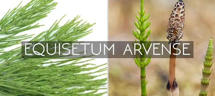
Equisetum Arvense (Field Horsetail)
Strobilus Of Equisetum
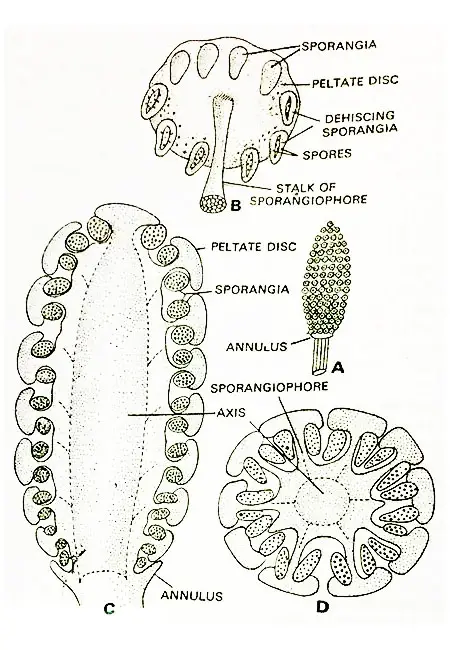
Equisetum arvense (Field Horsetail) consists of strobilus, each strobilus has a thick axis, having several whorls of densely crowded peltate appendages situated on the sporangiophores.
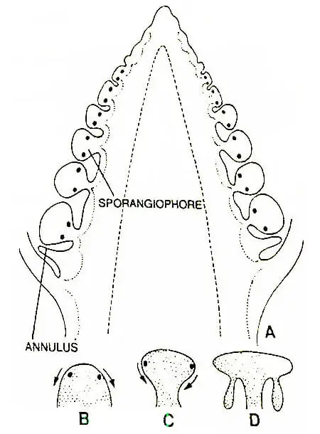
The sporangiophores are arranged in whorls. Usually, each such whorl is composed of twenty sporangiophores or so. In many cases, immediately below the sporangiophores, the central axis of the strobilus bears a small ring-like outgrowth, known as the annulus.
Each sporangiophore is composed of a single slender, stalk of which the terminal end is expanded into a flattened peltate disc situated at right angles to the stalk.
The peltate disc is usually hexagonal in outline in its surface view. This hexagonal appearance of the discs is attained due to the mutual pressure of the discs of the crowded sporangiophores situated on the strobilus.
Each peltate disc bears a ring of 5 to 10 isolated sporangia on its underside. Each sporangium is elongated and sac-like. On their maturity, the sporangia open by longitudinal slits for dispersal of the spores.
Development Of Sporangium
The development of the sporangia of Equisetum is eusporangiate type. This means, that they are not entirely derived from a single sporangial initial.
According to Bower (1894), all the sporogenous tissue in a sporangium (special enclosure structure which is responsible for the production as well as storage of spores) is developed from a single superficial cell of the young sporangiophore.
The development of the sporangia begins as the strobilus ceases to grow in length.
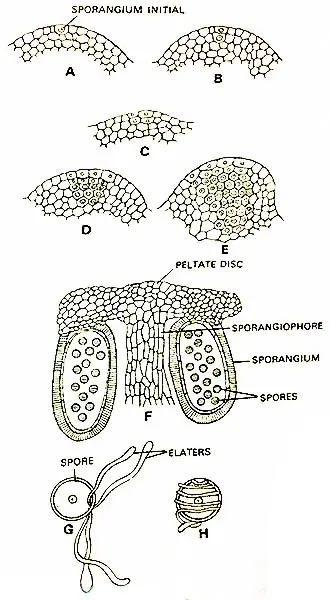
The young sporangiophores are found in the form of convex outgrowths in acropetal succession on the conical axis of the strobilus.
At the beginning of the development, the sporangiophore is hemispherical, but later on, this soon becomes constricted at its base developing a short stalk and sporangia bearing peltate disc at its apical end.
The development of the sporangium begins from a single superficial cell known as sporangial initial situated in the peripheral region of the young hemispherical sporangiophore.
The center of the tip of the young sporangiophore increases in size actively and the sporangial initials are shifted to its lateral sides.
Sooner or later, because of further development, the outer edge of the sporangiophore becomes inverted and faces the axis, this way the sporangial initials have also changed their position, and now facing the axis.
At first, the sporangial initial divides periclinally forming the two daughter cells. The daughter cell gives rise to the sporogenous tissue by repeated division, whereas the outer cell gives rise to both the sporogenous tissue and the portion of the jacket of the sporangium radial to the sporogenous tissue.
The remaining portion of the jacket layer is developed from the cells laterally placed to the original initial. Ultimately the jacket layer becomes multilayered.
The innermost layer is the tapetum and supplies nourishment to the developing spores.
During the development of the spores, the tapetum and all but two layers of the jacket become disintegrated.
This way, the mature sporangium is surrounded by a two-layered jacket. The cells of the outermost layer of the jacket are spirally thickened. During the development, the cells of the sporogenous tissue become separated and act as spore-mother cells.
About one-third of the mother cells disintegrate. The disintegration of the mother cells, tapetum, and the other jacket layers, together form a plasmodial fluid in which the remaining two-thirds of spore mother cells float about.
Ultimately the spore mother cells undergo usual meiotic division developing the spore tetrads. Each tetrad is composed of four haploid spores.
Structure Of Sporangium
Each mature sporangium is elongated, sac-like, and rounded at its apex. on maturity, it consists of a single-layered intact layer. By now, the rest of the jacket layers are aborted. The sporangium contains within it the mature spore all alike.
The Equisetum is homosporous. The sporangia are attached to the underside of the hexagonal peltate disc in a ring. Each sporangiophore bears 5-10 sporangia.
Dehiscence Of Sporangium
On the maturation of the spores within the sporangium, it bursts by a longitudinal slit. Prior to the opening of the sporangia, the internodes of the strobilus elongate separating the sporangiophores quite apart from each other.
The walls of the cells of the jacket layer are spirally thickened they shrink and lose water and the longitudinal slit develops on the sporangium for the release of the spores.
The Spores And Work Of Campbell
According to Beer (1909), the spore (spores are defined as agents of sexual/asexual reproduction which are adapted in later stages of the life cycle for dispersal or survival, especially in plants, bacteria, fungi, and algae) secretes a wall around it, composed of four concentric layers.
The outermost layer is known as the exosporium and the innermost endosporium. Outside the exosporium, there is a delicate cuticle called the middle layer.
On the extreme outside of the spore there lies the epispore which later on develops into four strips that get separated from each other from the rest of the wall but or one common point of attachment.
These strips are elaters. The strips or elaters remain coiled around the spore unless and until it is not disseminated from the sporangium. The elaters are hygroscopic.
They remain coiled and uncoiled around the spore due to the changed conditions of the moisture of the air. When the air is moist, they remain coiled around the spore and as soon as the air is dry the elaters are uncoiled.
The elaters of Equisetum do not resemble in any respect to elaters of Bryophyta. The ends of the spiral elaters are spoon-like. Each spore contains a centrally located nucleus and many small chloroplasts. The spores remain viable only for a few days after they are released from the sporangium.
Function Of Elaters
The function of elaters is not very certain, but it is assumed their expansion may help in the dehiscence of the sporangium. It is also thought that their hygroscopic movements may assist in the dispersal of the spores.
According to Buchtien due to the presence of elaters, the spores are entangled together and thus dispersed in groups so the Prothalli are found in close vicinity. According to Goebel, the elaters help in fastening the spore to the Substratum.
Germination Of Spore And Development Of Prothallus (Gametophyte)
Usually, the spores germinate in the clayey soil and mud along the banks of the rivers and streams. As already mentioned, the spores contain chloroplasts and therefore they can survive only for a few days after their liberation from the sporangium.
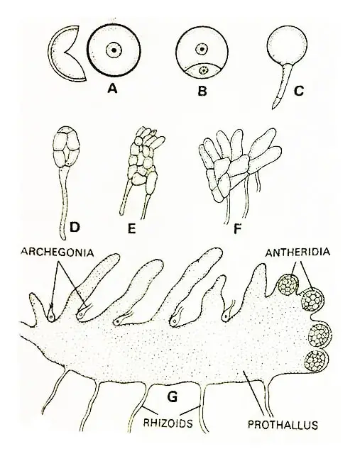
Prior to germination, the spore increases to some extent in its size. Within 10 to 12 hours, the first cell division in the development of the gametophyte (Gametophyte definition: the sexual phase in the alternation of generations of plants and certain algae) takes place, after the spore is sown, the two daughter cells result in one small and the other large in size.
The small cell develops into the first rhizoid and the large one gives rise to the remainder of the thallus (Campbell, 1918 | Kashyap, 1914 | Walker, 1931). The large cell divides irregularly several times giving rise to one to several celled thick cushion-like tissue.
This cushion-like structure bears numerous rhizoids on its towel surface. This cushioned is known as gametophyte or prothallus. Upon this cushion-like massive structure, the green lobes or branched are found.
The prothallus is elongated and lobed. The cells of the lower portion of the cushion are colorless whereas those of the portion bear chloroplasts and are green.
The entire margin of the prothallus is meristematic. When the growth of the entire margin is uniform the prothallus becomes quite smooth and circular.
Whereas on the other hand when growth is more active on one side or the other, the irregularly lobed prothallus has resulted.
The upright green lobes arising from the upper side of the prothallus are densely crowded and almost cover the whole upper surface of the gametophyte.
In certain species the gametophytes are quite big in size, e.g., in E. debile Rox, the gametophyte is 3 centimeters in diameter (Kashyap, 1914), and two to three millimeters in thickness.
In the majority of the cases, the gametophytes are less than a centimeter in size. The gametophytes are long-lived. According to Walker (1921), it has been reported on the basis of cultural experiments that they can survive for more than two years. The large gametophytes survive for a long period, whereas small prothalli die soon.
Nature Of Prothalli
There are two types of prothalli-the small male prothalli bear only antheridia whereas the large hermaphrodite prothalli bear both the antheridia and archegonia. whether Equisetum is monoecious or dioecious is a matter of controversy.
Kashyap (1917)
According to Kashyap’s research, in E. debile when spores are sown thickly, the prothalli, remain small in size and bear only one type of sex organ.
On the other hand, if the spores are shown quite separate from each other the prothalli become large in size and bear both types of sex organs, i.e., antheridia and archegonia. In such cases, however, the archegonia develop first and thereafter the antheridia.
Walker (1921)
According to Walker (1921), E. laevigatum is a monoecious species. In Equisetum arvense, under unfavorable conditions, the prothalli remain small in size, and only antheridia are developed, upon them. In favorable conditions, half of the prothalli are developed as males and the remaining half as females.
According to Walker, the normal prothalli are monoecious, which develop archegonia first and the antheridia later on. He also stated that the prothalli developing in crowded conditions remain small and dioecious.
In Equisetum arvense, the normal gametophytes produce archegonia at first and thereafter antheridia. In conclusion, the Equisetum arvense is monoecious. This view has been supported by several eminent bryologists.
Joyet Lavergne (1931)
According to Joyet Lavergne (1931), morphologically the spores are alike, but physiologically they are different, and in the same lot some of the spores ate and some are female, so according to him, there is internal heterospory.
Development Of Archegonium In Equisetum Arvense
If favorable conditions prevail, the first sex organs appear when the normally separated gametophytes are 30 to 40 days old. The sex organs appear only when they are differentiated into two green lobed regions and the lower massive tissue.
Normally archegonia first develop in the meristematic margin of the prothallus. They are situated among the upright green lobes.
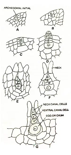
The archegonial initial is a superficial cell situated in the meristematic margin of the prothallus.
The archegonial initial divides first by a periclinal wall giving rise to two daughter cells.
The outer daughter cell is known as the primary cover cell and the inner is known as the mother cell of the central cell.
The primary cover cell divides further producing four primary neck cells. The four primary neck cells divide transversely producing an archegonial neck with three or four cells in height.
The central cell divides by a transverse wall producing a primary neck canal cell and a primary venter cell.
In some species, the primary neck cell rarely divides, whereas in other species it divides regularly producing two boot-shaped neck canal cells.
In almost all the species, the primary venter canal cell divides by an asymmetrical transverse division producing a venter cell and an egg. Lastly, all the axial cells situated in the archegonium are disintegrated but the egg.
Structure Of Archegonium
An archegonium (the female reproductive organ in plants) consists of a sunken base in the prothallus and a projecting neck. The archegonial neck is composed of four vertical rows of cells. Each row is two to four cells in height.
The terminal cells of the archegonial neck are quite long, which become separated and curved outward on their maturity. The axial row of the cells situated within the archegonium consists of three or four cells-the egg, the venter canal cell, and or two neck canal cells.
When there are two neck canal cells, they are boot-shaped, e.g., E. debile. In the last stage of the development of archegonium the axial cells are disintegrated but the egg.
Development Of Antheridium In Equisetum Arvense
In Equisetum arvense (Field Horsetail) as soon as the formation of archegonia is over the antheridia are formed on the same prothalli.
At the time of the development of antheridia, the growth of the upright lobes on the prothallus is stopped, and the prothallus turns and expands upwards.
The antheridia develop in these expanded and turned portions of the prothalli.
On the other hand, in starved, crowded prothalli, only antheridia are developed. These prothalli may also be called the male prothalli. Usually, the antheridia are of two types
- The embedded type
- The projecting type
The former type usually develops on the massive tissue of normal gametophytes and the latter type of antheridia is developed in the meristematic margin of the starved crowded and dwarfed male prothalli.
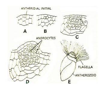
A superficial cell of the meristematic margin of the prothallus acts as an antheridial initial. The first division of the initial is periclinal.
This way, two daughter cells are formed. The outer daughter cell is known as the jacket initial and the cell is known as the primary androgonial cell.
The jacket initial divides rise to a single-layered jacket. Sometimes there lies a triangular opercular cell at the apex jacket layer.
The primary androgonial cell divides repeatedly giving rise to a group of cells.
Ultimately, the androcyte mother cells are formed. Near the nucleus of each mother cell, two blepharoplasts appear.
Now, each androcyte mother cell divides and gives rise to two each androcyte each possesses a blepharoplasty.
Each androcyte metamorophoses into a spirally coiled multi-flagellate antherozoid.
In the beginning, the rounded nucleus and the blepharoplast of the androcyte become elongated. The blepharoplast is composed of many granules. Later on, all the granules become united.
The nucleus elongates continuously and ultimately becomes a crescent-shaped. In the same way, the blepharoplasty also becomes spirally elongated.
Later on, the posterior end of the nucleus becomes expansive and flattened. A large vacuole is developed within androcyte and the cytoplasm becomes foamy and coarse.
Structure Of Antherozoid
The antherozoids (they are defined as male gametes found in a haploid structure called antheridium) are quite large in size. Each antherozoid is spirally coiled.
The posterior part of the body of the antherozoids is thick and flat. The anterior part is conical and bears numerous flagella. The body is almost entirely derived from the nucleus of the androcyte
The numerous flagella at the anterior end of the antherozoid originate from the cytoplasm of the mother cell. The flagella are attached to the blepharoplast, which also lies at the anterior end.
Fertilization
On the maturity of the archegonium, the neck canal cells and the venter canal cell are disorganized. The antherozoids swim down the neck canal and approach the egg.
Several antherozoids travel down the neck canal but only one of them penetrates the egg. In E. debile, Sethi (1928) observed that the complete antherozoid enters the egg, and fertilization is effected by forming a diploid oospore (2x).
Unlike other pteridophytes, usually more than one archegonia are fertilized and several sporophytes are developed on the same prothallus. Usually, 8 to 10 develop on a single prothallus, e.g., E. debile. Kashyap (1914) has recorded as many as 15 sporophytes on a single gametophyte in E. debile.
Development Of Embryo (Young Sporophyte)
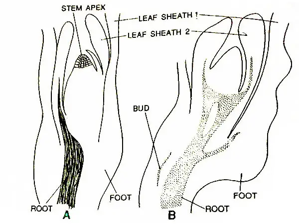
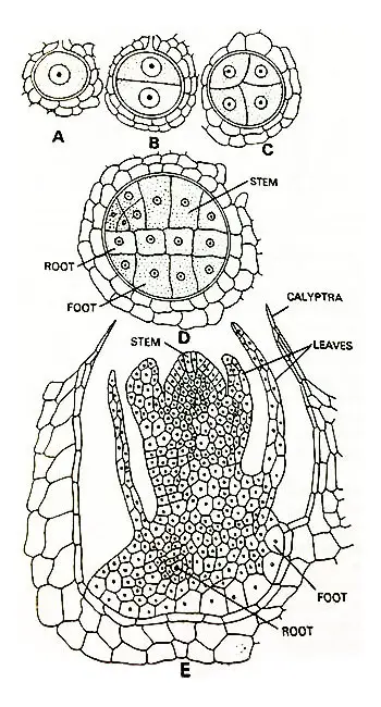
As supported by Campbell (1918, 1928), Jeffery (1899), and Walker (1921), usually the first division of the oospore is transverse, giving to two cells-the upper one is epibasal and the lower one the hypobasal.
Here in the case both the halves of the oospore take part in the development of the embryo proper and no suspensor is developed like that of Selaginella.
The next division takes place at a right angle to the first and the quadrant stage of the embryo is attained.
According to Sadebeck (1878) in Equisetum (Horsetail), the first division of the oospore is followed by two successive divisions at right angles to each other giving rise to eight cells.
According to Campbell (1921), in E. debile the entire hypobasal half develops into the foot.
Whereas in E. arvense (Sadebeck, 1878) the hypobasal half of the embryo gives rise to both foot and root.
Early in the development of E. arvense the epibasal cell divides by three intersecting walls in such a way that a tetrahedral apical cell of the stem is differentiated.
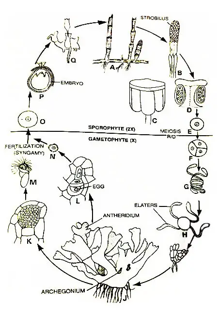
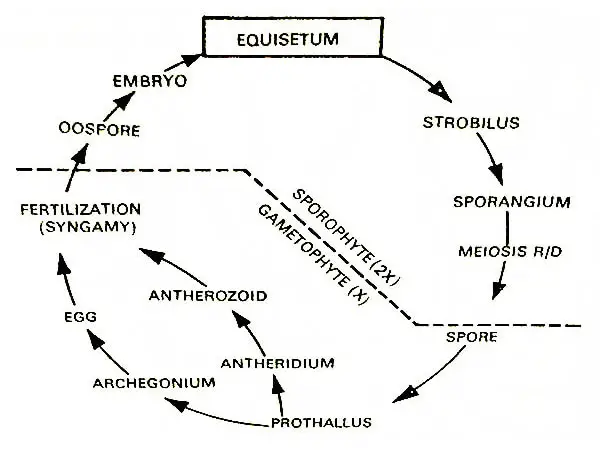
Graphical Life Cycle Of Equisetum
The three cells, lateral to this apical cell are also differentiated, which develop in the three leaves of the first leaf sheath.
Sometimes, there may develop two or four leaves instead of three. The so-developed leaves are quite small and scale-like.
In Equisetum debile, where the entire hypobasal half develops into the foot, a superficial cell of the epibasal half lying near the hypobasal half develops into the apical cell of the root.
The portions of the stem and root of the embryo grow and elongate quite rapidly.
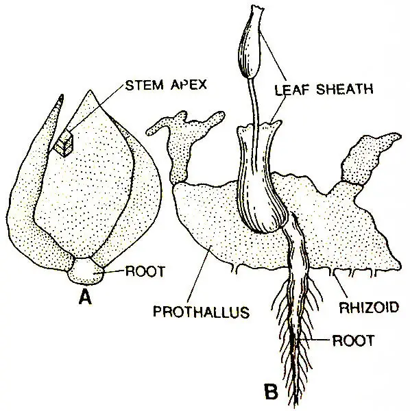
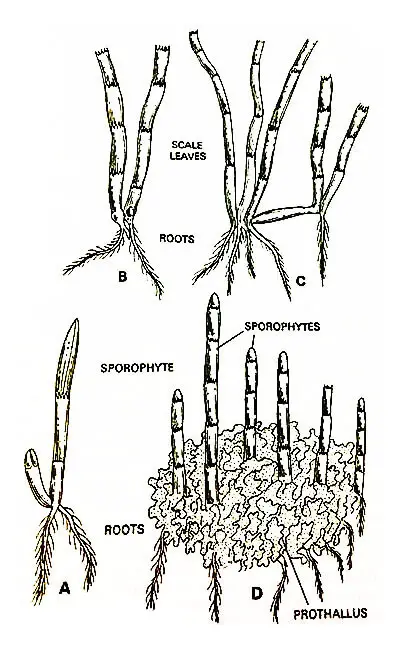
The stem portion of the embryo elongates and bursts through the neck of the archegonium, forming the calyptra and growing upright.
On the other hand, the root grows downward of the gametophyte and reaches the soil. Soon after, the stem differentiates into nodes and internodes.
Generally, a whorl of three leaves, or so is found at each node. The stem grows upward unless and until 10 to 15 nodes and internodes are developed.
Thereafter a secondary branch arises from the base of the primary branch. This branch bears four or five leaves at each node.
The growth of the secondary branch is also limited and reaches probably the same height as that of the primary branch. More vigorous branches arise successively from the base of the primary stem.
The third or four branch turns downward into the soil and becomes the rhizome of the (Horsetail) Equisetum plant.
According to Campbell (1928), the origin of the secondary branch is endogenous and it arises from the primary root. The secondary branch possesses adventitious roots.
Several secondary branches of Limited growth arise successively from the young sporophyte. All such branches are endogenous in their origin.
From the point of view of its anatomy, the primary stem is solid and carinal canals which develop later on in the adult stem.
However, the xylem is better developed in the primary stem. The base of the seedling bears protostele, but later on, the stem bears siphonostele at the level of the first branch.
Economic Importance
Some of the species of equisetum (Horsetail) are used in the preparation of the indigenous medicines
- Equisetum arvense is used as diuretic
- Equisetum debile is used as cooling medicine.
- Equisetum arvense is used as a natural, herbal, health supplement.


Good Work on your latest blog post. I have been checking for additional data on this topic for quite some time now.