Bacteria Guide | The Life Cycle of Bacteria 2024
Table of Contents
Bacteria are those organisms, which can be found throughout the biosphere. They are the simplest and smallest (< 1 µm — 50 µm width/diameter) organisms.
They belong to the group prokaryotes (Gr. Pro, before; karyote, nucleus), which can be studied even by a light microscope. Here I have described in detail the life cycle of bacteria in order to understand the complexities this micro-organism has.
What is Bacteria or Give Bacteria Definition?
According to the microbiology definition of bacteria, they are one-celled and are classified as microscopic organisms found throughout the whole environment.
In soil, in air, in water (even in boiling water), and even in your own body. For example, the bacteria Escherichia coli. Bacteria play an essential role in our lives and hold economic importance in both agriculture and industry.
E Coli Morphology
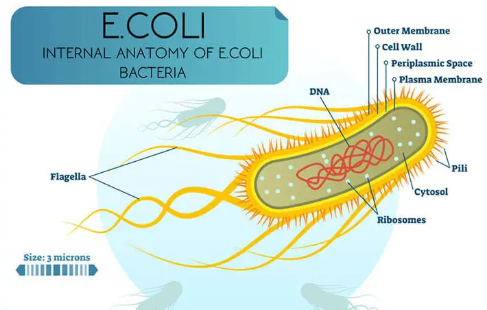
Here is the external and internal structure of E. coli.
History of Bacteria Discovery
Antonie Van Leeuwenhoek (Father Of Microbiology)
Anton von Leeuwenhoek discovered bacteria in 1675 without any major understanding of the organism and give them the name animalcules.
Pasteur And Koch
But French Microbiologists, Pasteur, and Koch had the honor to make a major contribution in the field from 1870 to 1976.
They were able to identify several bacteria morphologically and physiologically.
Koch was able to demonstrate that bacteria can cause diseases such as Anthrax tuberculosis. This understanding leads them to suggest “The Germ Theory of Disease”. The idea was soon accepted and studies on it as a subject were established.
Thomas Jonathan Burrill (Father Of Mycology)
Originally the idea was restricted to animals only.
T. J. Burrill was the first to demonstrate that even plants could develop diseases caused by bacteria.
A lot of information about the bacterial kingdom has been added to human knowledge since then.
Classification Of Bacteria
It is a traditional approach that classification is based on morphological characters. But in bacterial cases, because of their simple morphology, they cannot be divided into orders, families, genera, and species on the basis of their structure alone.
Thus, their classification studies are carried out in the field of their biochemistry, physiology, and cultural characteristics.
We have a number of classifications of bacteria but the most accepted classification is believed to be the classification carried out by Bergey (1957). Some other classifications are also quoted for a better understanding of the subject.
Erwin Frink Smith
Erwin F. Smith, a pioneer worker in this field of bacterial classification accepted a system of classification proposed by Migula, which was based on the number and arrangement of flagella. A general outline of the classification is as follows.
- Bacterium included all atrichous rod-like forms.
- Pseudomonas included polar flagellated forms.
- Bacillus included peritrichous rod-like forms.
Smith in 1905, revised this system with slight modifications in the name of the divisions, thus the name
- Aplanobacter gave for atrichous, rod-like bacteria
- Bacterium for polar flagellate bacteria
- Bacillus for peritrichous bacteria
The commonly used and more widely accepted divisions of bacteria are,
- Bacterium or Bacillus included all rod-like and idly shaped forms.
- Coccus includes all-spherical bacteria.
- 3, Spirillum included spiral, comma-like, and variously curved bacteria.
Bergey’s Manual Of Determinative Bacteriology (1957)
A system of classification of Schizomycetes that is used by all bacteriologists and that has an international standing was presented by Berger in 1957 in “Bergey’s Manual of Determinative Bacteriology”.
It represents the collaborative efforts of over 100 best-qualified microbiologists of that time. It was established by the International Committee of Bacteriological nomenclature in 1947.
The system was based on the international nomenclature rules of viruses and bacteria. As outlined in Bergey’s manual all bacteria were divided into about 1500 species.
These all bacteria were differentiated from one another primarily on the basis of type of motility and morphological characters. They are divided into a total of 10 orders.
Order Pseudomonadales
Cells are rigid, spheroids or rod-shaped, straight, curved, or spiral some groups form trichomes; motile species have four flagella.
Chlamydobacteriales
In trichomes rod-like cells are present. They are often sheathed and iron hydroxide is deposited in the sheath. The flagella are sub-polar when present.
Hyphomicrobiales
Cells spheroidal or ovoid, connected on stalk or thread, on trichomes; exhibits budding and longitudinal fission; flagella polar when present.
Eubacteriales
Typically, eubacteriales have rod-like or unicellular spheroidal cells present on trichomes. They consist of a sheath or other accessory structures. In eubacteriales, the motile species have peritrichous flagella.
Order Actinomycetales
Cells branch and many species form mycelia and mold-like conidiophores and sporangiophores, polar flagellate, and sporangiospores in only one species.
Caryophanales
Trichomes are often very long, peritrichous flagella.
Beggiatales
Alga like trichome or coccoid cell, accumulate elemental S, gliding and oscillatory or rolling motion; no flagella.
Mycobacteriales
Cells coccoid, rod-like or fusiform, communal slim, fruiting bodies, cells flexuous, gliding motility in contact with the solid surface; no flagella.
Spirochaetales
Elongated, spiral cells rotatory and flexing motions and translator motility; no flagella.
Mycoplasmatale
Extremely pleomorphic and easily distorted cells without cell walls; complex life cycle; non-motile.
Table Of Comparative Study Of Bacteria By Different Workers
A comparative study of bacteria by different workers in this field is as follows:
| Plant Nutrients | Time Period | Non-Motile | Motile With Polar Flagella | Motile With Peritrichous Flagella |
|---|---|---|---|---|
| Migula | 1895 - 1900 | Bacterium | Pseudomonas | Bacillus |
| Lehmann And Neumann | 1897 - 1927 | Non-spiral motile or non-motile bacterium | Sporing motile or non-motile | Bacillus |
| Smith | 1905 | Aplanobacter | Bacterium | Bacillus |
| Bergy | 1923 - 1939 | Phytomonas | Erwinia | |
| Dowson | 1939 | Pseudomonas | Pseudomonas And Xanthomonas | Bacterium |
| Bergey | 1948 | Pseudomonas | Pseudomonas And Xanthomonas | Bacterium |
Phylogenetic Relationship | Bacterial Phylogeny
The phylogenetic relationship of the bacteria is not certain. This uncertainty primarily becomes the cause of a long delay in the proper classification of this group.
According to one scheme, they were placed in the phylum Schizomycophyta in the Sub Kingdom Thallophyta, which includes all plants and does not form embryos during development.
According to another modern taxonomic system these organisms have been placed in the Kingdom Protista along with algae, fungi, and protozoan.
But the most modern understanding of the five Kingdom systems proposed by Robert Whittaker in 1969 placed them in the kingdom Monera along with blue-green algae, on the basis of their Prokaryotic nature.
According to one school of thought, the bacteria have been, descended from the blue-green algae, after becoming adopted to a saprophytic or parasitic lifestyle. This view has its general understanding based on the similarity of the cell structure of these two forms.
According to other investigators, the fact that many bacteria possess flagella indicates that these organisms descended from simple flagellated forms and perhaps these forms have also given, rise to the green algae.
There are others who believe that the heterotrophic bacteria of today have evolved from autotrophic ones. Well, according to these workers, before the appearance of any chlorophyll-containing plants the autotrophic bacteria may have appeared.
It is also suggested that different groups of bacteria from different ancestors have descended independently.
Many others still believe that bacteria have given rise to other forms as being a terminal group in evolution or if they have not descended from the blue-green algae, perhaps the blue-green algae have descended from them.
Occurrence And Characteristics of Bacteria
Occurrence
They are omnipresent and found almost everywhere but they are in great abundance in tropical and temperate regions.
On one hand, they can be found in the snow at temperatures less than zero but on the other hand, they can also be found in hot springs with a temperature of 78°C.
What Are The Characteristics Of Bacteria?
The majority of them are unicellular but the filamentous and colonial forms are quite common. Usually, they are found embedded in the mucilaginous envelope.
Even when the bacteria are held together in chains each individual cell carries on its own metabolic process quite independently.
They may be devoid of chlorophyll and mostly heterotrophic in nutrition.
They produce asexually chiefly by fission while sexually they have three different modes like conjugation, transformation, and transduction.
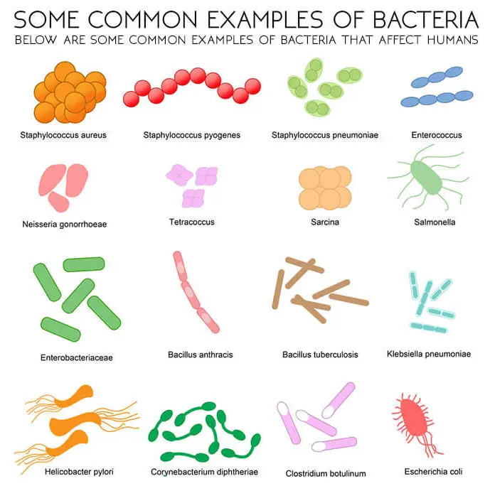
Before going towards the morphology of bacteria, here we have a few examples of bacteria that affect humans. The chart is given below.
General Morphological Characters Of Bacteria
Size And Shape Of Bacteria
The bacterial cell varies greatly in their size. An average bacterial cell measures 3µ long and 1µ broad whereas an average spherical cell is 1µ in diameter.
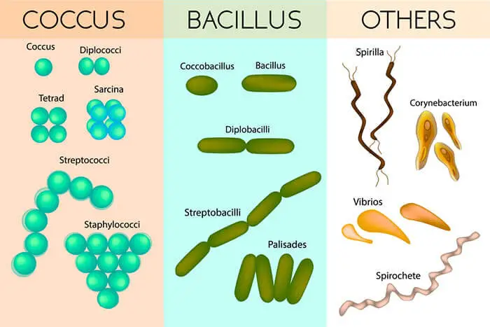
The average volume of a bacterial cell is 1 cubic micron. It has various forms but the abundant three basic forms of bacteria are Coccus, Bacillus, and Spirillum.
Coccus
They are rounded or spherical. They are the smallest forms of bacteria. The cell may either separate from each other or may remain joined ‘together after the division to form groups of two (Diplococcus) or four (Micrococcus tetra-genus) or it may be a chain of cells (Streptococcus). The average size of coccus bacteria ranges in diameter from 0.5µ to 1.25µ.
Bacillus
They are straight rod-like and possess rod-like, kidneys or elongated cells. They vary greatly in their length and diameter ranging from 0.6µ to 1.2µ long and 0.5µ to 0.7µ wide to 3.8µ long and 1 to 1.2µ wide.
Spirillum
They are spiral or curved forms. In this case, the cells vary in size from 1.5µ to 4µ long and 0.2µ to 0.4µ wide in vibrio and up to 50µ long in spirillum.
Pleomorphism
Under favorable conditions of growth and development, the shape of one species remains constant, whereas under unfavorable conditions some bacteria such as nitrogen-fixing bacteria change into at least three different forms.
Besides the original structure of the parent type, many bacteria show a mixture of several integrating forms in young cultures. This phenomenon is known as pleomorphism.
When treated with an antibiotic, attacked by T4 bacteriophage or cold shook they sometimes develop soft protoplasmic forms, which are known as large bodies or L-forms. If transferred back to Normal conditions their form also reverts back to normal shape.
Structure Of Bacterial Cell (Bacteria Anatomy)
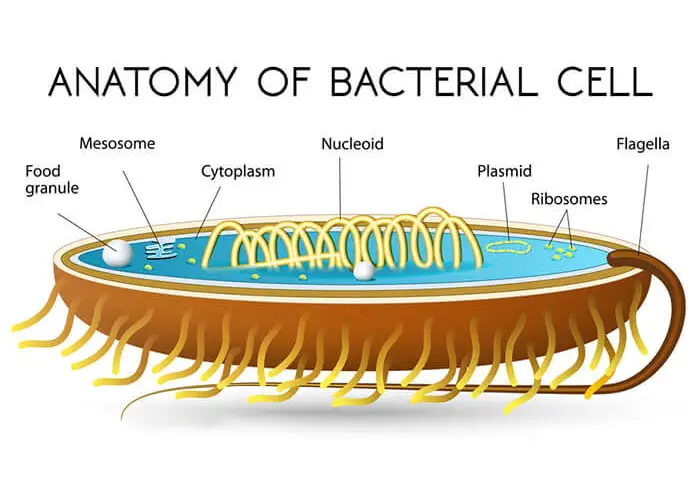
The internal structure of bacteria cells contains vacuoles, ribosomes, and granules of stored food. It also contains granules of glycogen, proteins, and fats but lacks an endoplasmic reticulum.
Mitochondria are absent. The Mitochondria are defined as rod-shaped structures or organelles that are classified as power generators/powerhouse of the cell, which converts nutrients and oxygen into (ATP) adenosine triphosphate)
Enzymes found in the mitochondria are localized on or near the cell wall. Water is an important constituent about 90 % of cell is water. The movement of materials in and out of the cell is regulated by a cell membrane.
Structurally the cytoplasm of a bacterial cell is similar to the cytoplasm found in the living cells of more complex organisms. The membrane plasmalemma invaginates to become a complex structure known as mesosomes.
It is believed that mesosomes have an active role in cell division by wall synthesis and in the secretions of extra-cellular substances. The mode of cell division is amitotic.
Bacteria Nucleus
A nuclear membrane is not present. The prokaryotic nuclear region appears as an electron translucent area and can be shown to contain very fine fibrils in the electron microscope. These fibrils are molecular strands of DNA.
The DNA is a centrally restricted area of the cell. It has been demonstrated for some species of bacteria that the DNA is about 1200 microns long. i.e. more than 500 times long as the bacterial cell that contains it. The adjustment is made by supper coiling of the single circular chromosome.
Bacteria Cell Wall
A definite thin cell wall consisting of a single layer is present. The cell walls become gelatinous in some cases and appear like a sheath or capsule which holds cells together to form colonies. The cell wall accounts for 20% of the dry weight of the cell. It gives shape and firmness to the cell.
The chemical nature of the wall demonstrates that it is made up of highly complex, which consists of proteins, polysaccharides, and lipids.
The complexity of the wall is generalized into two categories i.e., Gram-positive bacteria and Gram-negative bacteria.
Define Flagellation And Its Types
Bacteria bear thin elongated thread-like structures called flagella (singular-flagellum), which help in their locomotion. There are different forms of bacteria that bear flagella. Almost all the spirilla, most of the bacilli, and some of the cocci are flagellated.
Flagella are the motile organs, which give a speed of about 50mµ per second, although they are much smaller than the eukaryotic flagella. They are about 120 to 180°A in diameter. Bacteria flagella consist essentially of a protein of a pure protein called “flagellin”.
The flagella arise from small basal granules as they are fixed onto the protoplast. The flagella may consist of either a simple filament onto the protoplast and arise from small basal granules, the blepharoplasts situated on the outside of the cytoplasm.
Types Of Flagella Arrangement
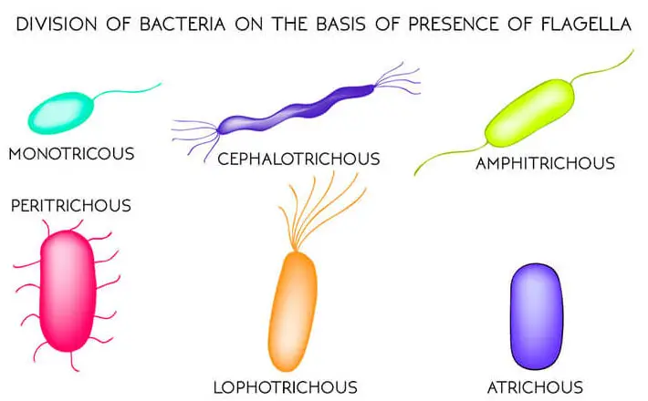
Here bacteria are divided into different types according to the presence of flagella.
Atrichous Bacteria
Those bacteria in which flagella are absent are termed Atrichous.
Define Monotrichous
Those bacteria which have only one polar flagellum are known as Monotrichous e.g. Vibrio.
Lophotrichous Bacteria
Another type is Lophotrichous. Now, these are types of bacteria that have a group of flagella present at one end. For example. Spirillum.
Amphitrichous Bacteria
On the other hand, those bacterial forms which have a group of flagella at both ends are named Amphitrichous.
Peritrichous Bacteria
Those types of bacteria which have flagella uniformly distributed all over the body are known as Peritrichous. e.g. Salmonella.
What Is The Structure Of A Flagella
The bacterial flagella is a non-flexible structure consisting of a single filament composed of many subunits of the protein flagellin.
The filament of the bacterial flagellum is attached to the cell by a hook and a basal body, which has a set of rings that attach to the cytoplasmic and a rod that passes through the rings to anchor the filament to the cell.
In Gram-negative bacteria, the basal body has two sets of rings, with each set containing two rings. The two rings which are attached to the cytoplasmic membrane are designated S and M and the two rings that attach to the outer membrane of the envelope are designed as L and P.
But the Gram-positive bacteria have only one set of rings and this set is attached to the cytoplasmic membrane. These rings are also designated as S’ and M. The hook structure attaches the filament of the bacterial flagellum to the rod of the basal body.
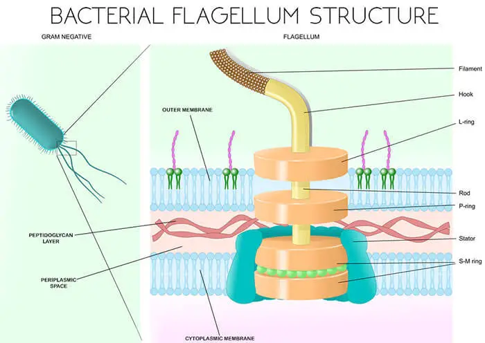
The structure of the bacterial flagellum allows it to spin like a propeller, with the basil body acting like a motor to rotate the filament and thereby propel the bacterial cell.
Rotation of the flagellum requires energy, which is supplied by the proton gradient across the cytoplasmic membrane. Approximately 256 protons must cross the cytoplasmic membrane to power a single rotation of the flagellum.
The flagellum can rotate at speed of up to 1,200 revolutions per minute, thus enabling the bacterial cell to move at speed of 100 µm/second (0.0002 mile/hour).
Considering that a typical bacterial cell has a maximum length of 2 µm, a rapidly swimming bacterial cell can move 50 times its body length per second – or in relative terms, twice as fast as a cheetah.
The Life Cycle Of Bacteria | Growth In Bacteria
Cell Growth
If conditions are favorable, cell division is normally followed by a period of elongation and growth. Under favorable conditions of moisture, nutrition, pH, and temperature some bacteria may double in about 20 minutes.
Different types of bacterial species show a variety of growth shape curves depending upon the generation time and the maximum population attainable under the growth conditions that prevail.
During the period of one to several, hours there may be a lag phase in which there is little or no increase in cell number. During the first part of the lag phase the cells adoption to the new environment.
Growing bacteria continue to do so at regular intervals until the maximum growth that can be supported by their environment is appreciated. This period of rapid cell division is known as the Logarithmic Growth Phase.
The stationary phase occurs when rapid growth is halted by the depletion of nutrition, accumulation of waste products, or other factors. The cells will eventually die if they are not transformed into the new environment which is capable of supporting the continuing support.
The Life Cycle Of Bacteria | Reproduction in Bacteria
In bacteria, both asexual and sexual reproduction is reported.
Asexual Reproduction
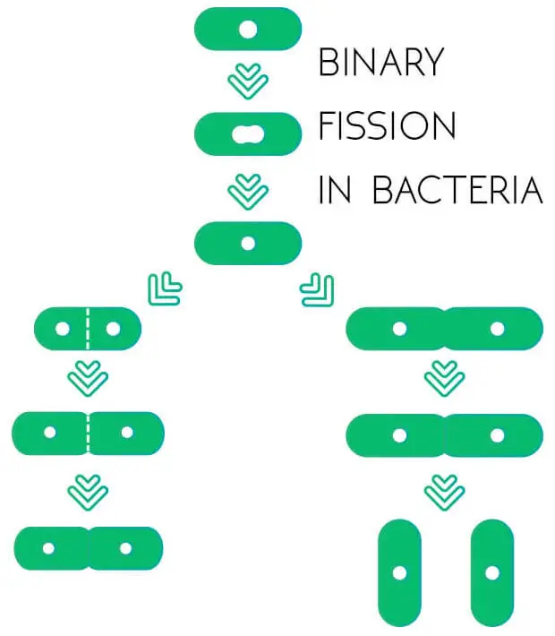
The common method is Binary Fission. Under ideal conditions of temperature, moisture, pH, and food bacteria show a rapid cell division after every 20 minutes.
If all the conditions remain optimum a man with simple knowledge can calculate the huge mass of bacteria accumulated within a few hours.
But practical knowledge proves this calculation to be wrong. In some cases, there may be an exhaustion of the food supply or an accumulation of waste products, which may be alcohol or some acid. Their accumulation retards the growth of the bacteria.
The division begins with the doubling of the DNA, which is followed by the division of all constituents distributed equally among the two daughter cells. The method is very fast and son their number increases proportionate to the prevailing conditions.
Sexual Division | Types Of Sexual Reproduction In Bacteria
Sexual reproduction is not a common phenomenon in bacteria. In general, understanding sexual reproduction involves an exchange of genetic material between two individuals.
In bacteria, at least three different modes of exchange of genetic material have been demonstrated. The list includes bacterial conjugation, transduction, and transformation.
Process Of Conjugation In Bacteria
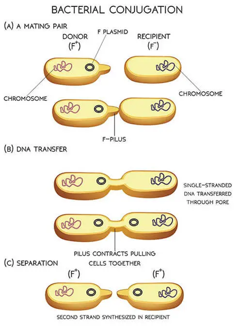
Conjugation is the mechanism by which genetic material is exchanged between the two bacteria through a cytoplasmic bridge.
The whole DNA is not involved but a plasmid known as a conjugative plasmid (F+ fertility+) forms a mating pair with bacteria that do not contain a conjugative plasmid(F-) by means of an F-pilus on the surface of the bacteria.
The pilus contracts pulling the two bacteria together and the DNA is transferred.
This phenomenon is best studied in the bacteria E. coli, the conjugative plasmids have been demonstrated to have the ability to transfer themselves between bacteria and in some cases, also to transfer pieces of chromosomal DNA.
It has been found that F plasmids and allied plasmids carry a group of genes called the Tra (Transfer) Genes, which encode all the proteins required to form a mating pair with another bacterium not containing it.
The DNA is then transferred from plasmid-containing bacteria called the donor or F+ bacteria to the recipient F– bacteria. The plasmid is large enough to contain 95 kb (kilo bite) in size.
The cell containing F plasmids (F+) also produces a structure known as F-pili on its surface, which are encoded by the tra genes.
These are proteinaceous, filamentous structures that attach to the surface of bacteria due to a phenomenon called surface exclusion. The F-pilus retracts on contact and thus pulls the two bacteria together.
Bacteria Conjugation Diagram
Transduction In Bacteria
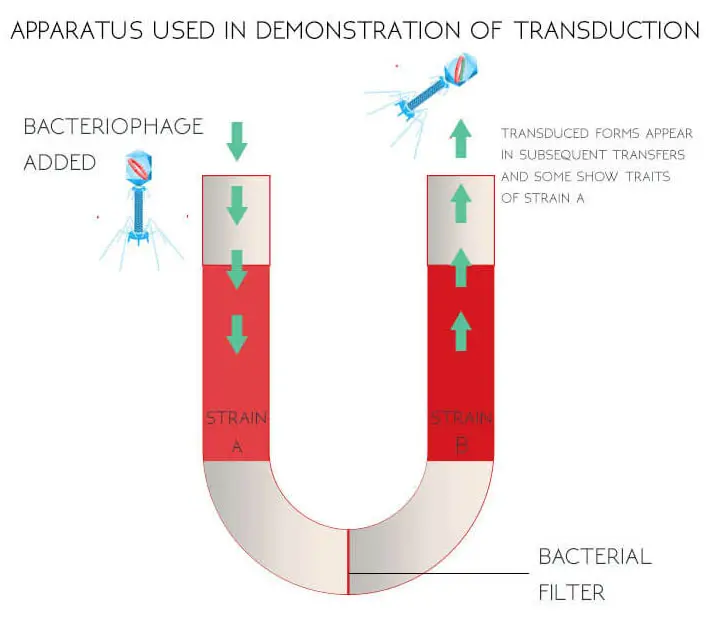
Transduction is a form of recombination in bacteria that involves the picking of a piece of DNA by phage from one bacterium called the donor which is then transferred to another bacterium called the recipient.
This phenomenon has been demonstrated in a wide range of bacteria and is thought to play a vital role in the transfer of genetic material between bacteria in nature.
Steps of Transduction in Bacteria
Transformation In A Bacteria
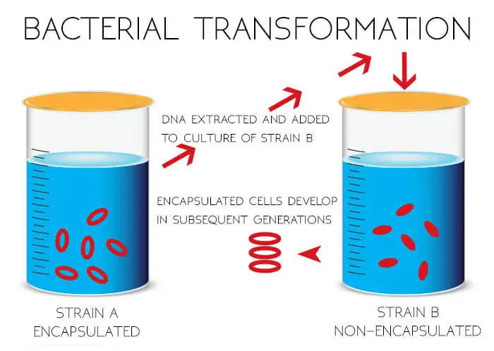
The phenomenon was discovered by Fred Griffith (1928). In Griffith’s bacterial transformation experiments, the free DNA molecule is transferred into recipient cells.
In this experiment, he found that DNA released from dead bacterial cells is genetically effective in very small amounts. In the laboratory, pure DNA extracts are used in transformation experiments.
Transformation typically involves only one trait, although several may be acquired independently in this process.

