Volvox Classification, Structure, Reproduction (2024 Guide)
Table of Contents
Volvox is a freshwater planktonic (free-floating) alga. There are about 20 species belonging to these genera. It is found in freshwater as green balls of a pinhead size.
They are just visible to the naked eyes, about 0.5 mm. in diameter. In the plant kingdom, it appears as the most beautiful and attractive object.
Globe Algae Volvox | The Chlorophyte Green Algae
The alga due to a specific swimming pattern is often referred to as, rolling alga. Its growth is frequently observed in temporary or permanent freshwater ponds, pools, ditches, and also in lakes.
The growth is abundant when temperature and organic matter are available in sufficient quantity. Its multiplication is so frequent and abundant that the water of ponds becomes green (water bloom).
The spring and rainy seasons are the usual periods of volvox’s active vegetative growth. With the onset of an unfavorable period (summer) the alga vanishes and passes an unfavorable period in form of the zygote. The volvox makes its own food by photosynthesis.
Volvox Classification
- Class: Chlorophyceae
- Order: Volvocales
- Sub-order: Chlamydomonadineae
- Family: Sphaerellaceae
- Genus: Volvox
The most common species of Volvox are:
- Volvox globator
- Volvox aureus
- Volvox prolificus
- Volvox rouseletti
- Volvox merelli
Volvox Under Microscope
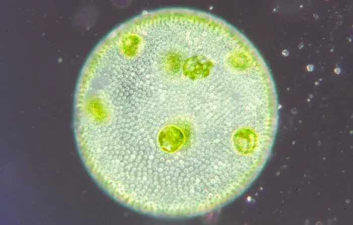
Below is the microscopic view of a colony of volvox:
Volvox Characteristics And Structure
Volvox Plant Body (The Gametophyte)
Volvox is a coenobial green-algae, {(the colony-plant body does not have a fixed number of cells e.g. Volvox aureus) (coenobium-plant body has a fixed number of cells, e.g., Pandorina moruma, number of cells are 4, 8, 16 or 32. Eudorina unicocca, number of cells 16, 32 or 64)}.
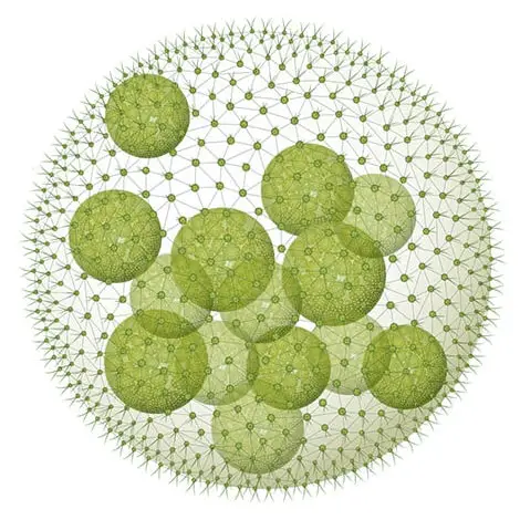
Among the motile forms, the coenobium of Volvox is the largest, highly differentiated, and well-evolved alga. Each coenobium is an ellipsoid or hollow sphere body with exactly marked delicate mucilage definite layer.
The interior part of coenobium is composed of diffluent (watery) mucilage, while cells are arranged in a single layer at the periphery.
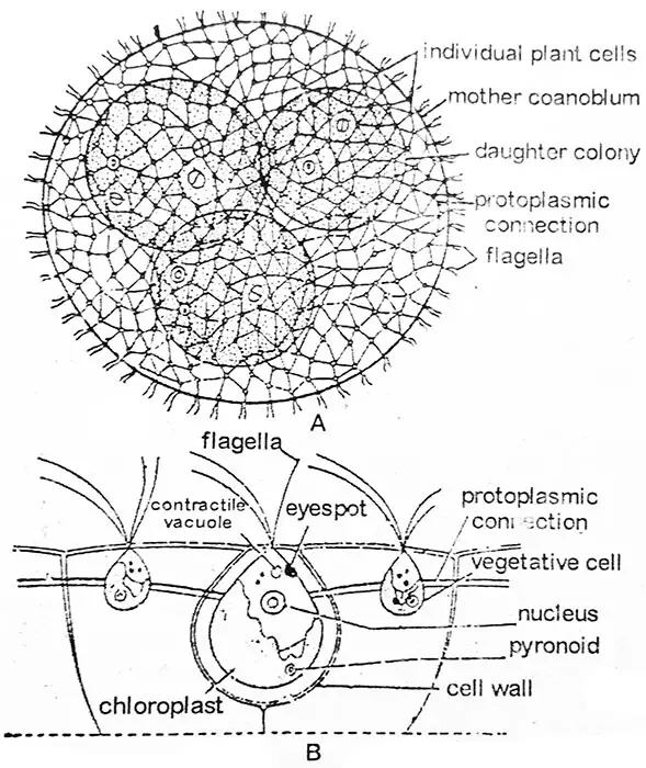
The movement of the colony is brought about by the joint action of the flagella of individual cells. Each coenobium has a definite anterior and a posterior end.
The coenobium shows polarity, it moves and rotates slowly, showing remarkable cooperation between the cells of the anterior and posterior end in the course of its movement.
Volvox is not an individual but an association of a number of similar cells, of which each function’s like an independent individual and carries out its own nutrition, respiration, and excretion and shows no cooperation between the cells in these functions.
The number of cells per coenobium varies e.g. 500-1000 in V. aureus, 1500-20,000 in V. globator, and even up to approximately 60,000 in V. rouseletti.
Volvox Cell Structure
Each individual cell has its identity. It is surrounded by its own large gelatinous, sheath, which may be conflicting with the sheaths of adjoining cells or may be distinct from one another.
Then they are distinct they are angular by mutual compression and are usually hexagonal in outline. Fig.,2.22. Thus, a considerable expanse of gelatinous material helps in separating one cell from the other cell.
In the majority of species, each cell is connected with its neighboring cells by a series of protoplasmic or cytoplasmic strands established during the course of cell divisions and the development of the colony.
The protoplasmic strands may be thin and delicate in V. aureus, round in V. globator, wedge-shaped in V. mononae, or may be absent as in V. tertius.
In outline, the individual cell of volvox resembles Chlamydomonas. Each cell has anteriorly inserted a pair of flagella of equal length. Both flagella are of whiplash-type.
The flagella project outside the surface of the coenobium into the surrounding water. Near the base of flagella two or more contractile vacuoles are present.
The protoplast is of different shapes depending upon the species. In V. tertius protoplast in V. aureus it is rounded and Chlamydomonas type, whereas in V. globator protoplast is a stellate type having diffused chloroplast and scattered contractile vacuoles.
Volvox Unicellular Or Multicellular
Vegetative cells of a young colony are green and alike in size and shape but in the older colonies, certain posterior region cells increase ten times; or more the size of the normal cell.
They develop numerous pyrenoids increase in size and behave as reproductive cells. They may be asexual or, sexual. In some cases, the same colony may bear both asexual and sexual cells.
In the anterior portion, the cells of the colony remain only vegetative and comparatively smaller. In the anterior region, cells bear a larger eyespot. So a colony consists of two types of cells: reproductive cells and somatic cells.
Volvox can serve as an example of the first step towards coordination and division of labor. A colony of Volvox can be regarded as a multicellular type composed of cells set apart for the performance of various functions. The cells performing different functions are,
- Vegetative cells are concerned with the manufacture of food and are involved in the locomotion,
- Asexual cells producing daughter colonies
- Sexual cells: producing eggs, and producing antheridium
Volvox Reproduction
In contrast to Chlamydomonas, the cells of the volvox colony show functional specialization. It reproduces both asexually and sexually.
At the beginning of the growing season (favorable conditions), the reproduction is asexual. It is for this reason that all the colonies collected at a time are either asexual or sexual.
Asexual Reproduction In Volvox
Asexual reproduction occurs at the beginning of the growing season during favorable conditions.
In the earlier stages, all the cells of a colony are alike but, later, a few cells in the posterior half of the colony store the food and increase in size.
These greatly enlarged cells are specialized asexual cells called gonidia (singular gonidium). Their number varies from two to fifty in a single coenobium.
Development Of Daughter Coenobium From Gonidium
Prior to the division, the gonidia are slightly pushed into the interior of the colony and can be distinguished as a row of vegetative cells by their larger size, rounded shape, absence of flagella and eyespot, prominent nucleus, several pyrenoids, and densely granular cytoplasm.
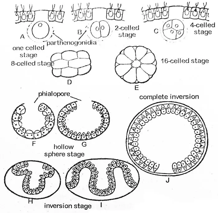
Each gonidium lies within a globular gelatinous sheath. The first division of the gonidial protoplast is longitudinal i.e. anterior-posterior plane of the coenobium.
The second division is also longitudinal and at a right angle to the first. Each of the daughter cells, thus formed, again divides length-wise so that an eight-cell plate is formed.
It gets curved with its concave surface facing outwards. This eight-cell stage is known as Plakea stage. Simultaneous longitudinal divisions of daughter cells continue for several cell generations (up to 14, 15, or 16 times in V. rouseletti).
At the sixteen-cell stage, the cells are arranged within the periphery of a hollow sphere, with a small opening, the phialopore towards the exterior of the parent coenobium.
At this stage, all the cells are naked and in contact with one another. Their anterior ends face the center of the sphere. With the progress of invagination, the phialopore greatly enlarges.
As the in-folding of a posterior portion (invagination) begins to push through phialopore. Its surrounding edges get curled backward which gradually slide down until the whole structure is inverted.
The phialopore which now shows a number of folds gradually becomes closed. The process of inversion requires about three to five hours. In some abnormal cases, the inversion does not take place at all as reported in V. minor.
The cells of the daughter coenobium now begin to separate from one another by the development of mucilaginous portions (cell wall). Each cell, finally, acquires a pair of flagella and a cell membrane.
The daughter colony (coenobium) is still retained within the parent cell wall which eventually develops into a mucilaginous membrane surrounding it.
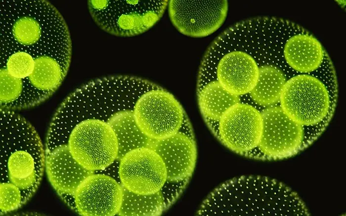
Several daughter coenobia may develop simultaneously in a parent colony. Thus, they may fill the hollow middle region of the parent colony.
The daughter coenobia is released with the death and decay of the parent colony. Sometimes the daughter colonies are not set free for a longer duration and develop granddaughter colonies.
Thus, as many as 2-4 generations of imprisoned daughter colonies may be seen in one original parent colony, especially in V. africanus.
Sexual Reproduction In Volvox
Volvox shows an advanced oogamous type of sexual reproduction which takes place by the formation of antheridia and oogonia. They may be formed on the same coenobium (monoecious) as in V. globator or on different coenobium (dioecious) as in V. aureus.
Monoecious species are protandrous (antheridia develop first) therefore, in such species fertilization will occur between the antherozoid and ovum of different plants.
It is quite interesting that sexual colonies are often devoid of asexually formed daughter colonies. In a coenobium, the cells destined to form sex organs are present in the posterior half. They are considered specialized cells.
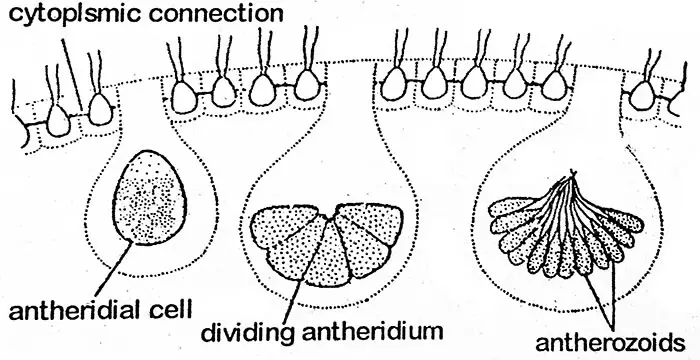
The sex organs (gametangia) are produced fewer in number. During the formation of gametangia, the cell becomes enlarged and rounded and discards the flagella but it remains connected with other cells by fine protoplasmic threads.
The male gametangium is called antheridium while the female oogonium. The protoplast of an antheridium undergoes repeated cell divisions in a way similar to that observed in the development of an asexual gonidial cell into a daughter colony (i.e. plakea stage).
Thus, a mass of small, naked, biflagellate, fusiform antherozoids 16 to 512 in number in an antheridium is formed.
These are grouped as flat plates except in V. aureus where antherozoids are seen in the asexual colonies. The plakea of antherozoids dissociates and liberates the antherozoids.
Antherozoid
Each antherozoid is a biflagellate, elongated, conical, or fusiform structure with a single nucleus and a small yellow-green or pale green chloroplast.
Oogonium
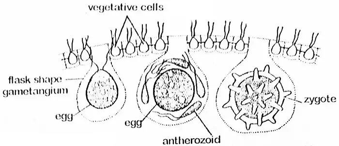
The oogonial cell enlarges considerably and discards its flagella and protoplasmic connections with the neighboring cells. The cell becomes rounded or flask-shaped with much of its portion projecting into the interior of the coenobium.
At this stage, it is called oogonium the entire portion of which is converted into a single spherical egg with a beak-like protrusion towards one side. Through this end, antherozoid enters the oogonium.
The egg (oosphere) contains a large centrally placed nucleus and a parietal chloroplast with pyrenoids. It is abundantly stored with reserve substances often absorbed from the neighboring cells through protoplasmic strands.
Fertilization
The antherozoids are liberated in groups at the time of fertilization and these remain intact till they reach the egg. The antherozoids are then, set free. Only one antherozoid fuses with the egg and results in the formation of an oospore.
The oospore subsequently secretes a three-layered smooth or spiny wall. It accumulates enough haematochrome (Red color pigment granules probably xanthophyll in nature) which gives it an orange-colored appearance. At this stage, it may be called a zygote.
Oospore And Its Germination
The outer wall and exospore may be smooth, (V. globator) or spiny (V. speematospaera). The middle layer is mesospore and the inner is the endospore. The zygote contains enough reserve food material and other inclusions.
Thus, the zygote is retained by the coenobium which can be liberated by the disintegration of the gelatinous matrix. After liberation, it settles down at the bottom of the pool and may remain viable for several years.

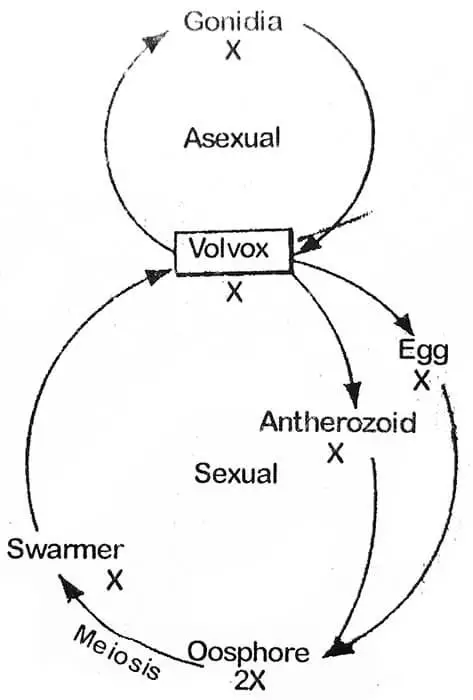
At the onset of favorable conditions, the zygote develops in different ways. In V. campensis the zygote nucleus divides meiotically and forms four nuclei, three of them degenerate and one survives: The survived nucleus accompanied by cytoplasmic contents escapes from the vesicle.
At this stage, it can be designated as a swarmer (large number or dense group, of insects, cells, etc.). It swims freely and divides and re-divides to form a new coenobium.
During germination outer two wall layers becomes gelatinous and the inner layer forms a vesicle which later on gets filled with the zygote protoplast.
In V. rouseletti and V. minor, the protoplast of the zygote is converted into a single zoospore which by further divisions forms a new coenobium.
Such coenobium consists of a smaller number of cells that reproduces asexually for the next six or more generations, every time increasing the number in the succeeding generations.
Volvox Life Cycle
The zygote is the only diploid phase in the life cycle of Volvox and therefore, the main plant body is haploid. That is why the zygote has to undergo reduction division during the formation of a new colony.

